 5 connective tissue pics legends.docx
- 文档编号:4906125
- 上传时间:2023-05-07
- 格式:DOCX
- 页数:23
- 大小:1,023.17KB
5 connective tissue pics legends.docx
《5 connective tissue pics legends.docx》由会员分享,可在线阅读,更多相关《5 connective tissue pics legends.docx(23页珍藏版)》请在冰点文库上搜索。
5connectivetissuepicslegends
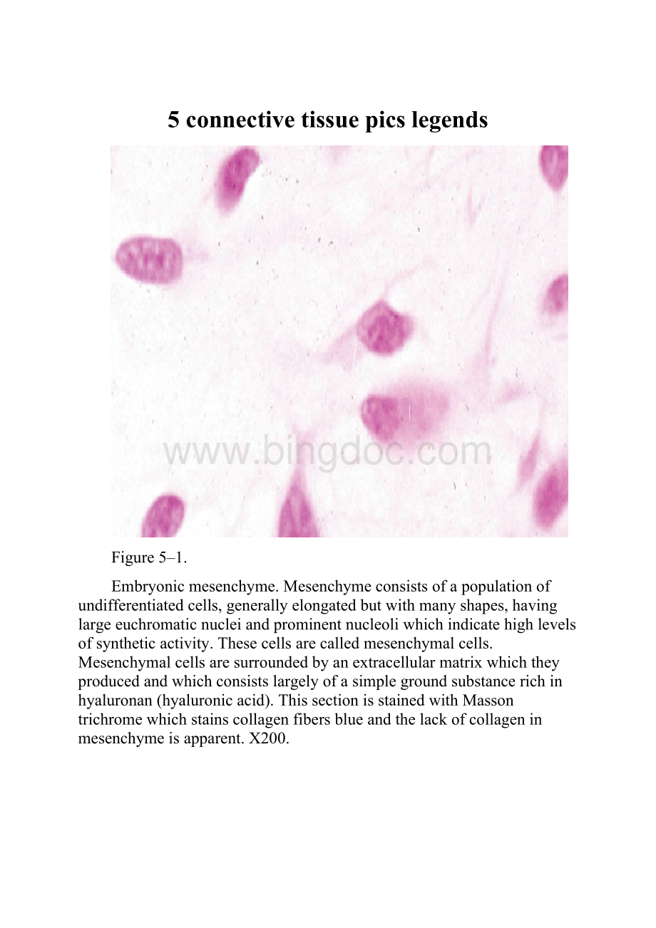
Figure5–1.
Embryonicmesenchyme.Mesenchymeconsistsofapopulationofundifferentiatedcells,generallyelongatedbutwithmanyshapes,havinglargeeuchromaticnucleiandprominentnucleoliwhichindicatehighlevelsofsyntheticactivity.Thesecellsarecalledmesenchymalcells.Mesenchymalcellsaresurroundedbyanextracellularmatrixwhichtheyproducedandwhichconsistslargelyofasimplegroundsubstancerichinhyaluronan(hyaluronicacid).ThissectionisstainedwithMassontrichromewhichstainscollagenfibersblueandthelackofcollageninmesenchymeisapparent.X200.
Figure5–2.
Lineagesofconnectivetissuecells.Thissimplifiedrepresentationoftheconnectivetissuecelllineageincludescellsfromthemultipotentialembryonicmesenchymecellsandhematopoieticstemcellsofbonemarrow.Dottedarrowsindicatethatoneormoreintermediatecelltypesexistbetweentheexamplesillustrated.Thecellsarenotdrawninproportiontoactualsizes,eg,adipocyte,megakaryocyte,andosteoclastcellsaresignificantlylargerthantheothercellsillustrated.
Figure5–3.
Fibroblasts.Connectivetissuewhereparallelbundlesofcollagenarebeingformed.(a):
Fibroblaststypicallyshowlargeactivenucleiandeosinophiliccytoplasmtaperingoffinbothdirectionsalongtheaxisofthenucleus,amorphologyusuallycalled“spindle—shaped.”Thenuclei(arrows)areclearlyseen,butthecytoplasmicprocessesresemblethecollagenbundles(C)thatfilltheextracellularmatrixandaredifficulttodistinguishinH&E—stainedsections.(b):
Bothactiveandquiescentfibroblastsmaysometimesbedistinguished,asinthissectionofdermis.Activefibroblastsarelargecellswithlarge,euchromaticnucleiandbasophiliccytoplasm,whereasinactivefibroblastorfibrocytesaresmallerwithlessprominent,heterochromaticnuclei.Theverybasophilicroundcellsin(b)areleukocytes.BothX400.H&E.
Figure5–4.
Macrophageultrastructure.CharacteristicfeaturesofmacrophagesseeninthisTEMofonesuchcellaretheprominentnucleus(N)andthenucleolus(Nu)andthenumeroussecondarylysosomes(L).Thearrowsindicatephagocyticvacuolesneartheprotrusionsandindentationsofthecellsurface.X10,000.
Figure5–5.
Mastcells.Mastcellsarecomponentsoflooseconnectivetissues,oftenlocatednearsmallbloodvessels(BV).(a):
Theyaretypicallyoval—shaped,withcytoplasmfilledwithstronglybasophilicgranules.X400.PT.(b):
Ultrastructurallymastcellsshowlittleelsearoundthenucleus(N)besidesthesecytoplasmicgranules(G),exceptforoccasionalmitochondria(M).ThegranulestainingintheTEMisheterogeneousandvariableinmastcellsfromdifferenttissues;athighermagnificationssomegranulesmayshowacharacteristicscroll—likesubstructure(inset)thatcontainspreformedmediatorssuchashistamineandproteoglycans.TheECMnearthismastcellincludeselasticfibers(E)andbundlesofcollagenfibers(C).
Figure5–6.
Mastcellsecretion.Mastcellsecretionistriggeredbyre—exposuretocertainantigensandallergens.MoleculesofIgEantibodyproducedinaninitialresponsetoanallergensuchaspollenorbeevenomareboundtosurfacereceptorsforIgE
(1),ofwhich300,000arepresentpermastcell.Whenasecondexposuretotheallergenoccurs,IgEmoleculesbindthisantigenandafewIgEreceptorsveryrapidlybecomecross—linked
(2).Thisactivatesadenylatecyclase,leadingtophosphorylationofspecificproteinsand(3)entryofCa2+andrapidexocytosisofsomegranules(4).Inaddition,phospholipasesactonspecificmembranephospholipids,leadingtoproductionandreleaseofleukotrienes(5).Thecomponentsreleasedfromgranules,aswellastheleukotrienes,areimmediatelyactiveinthelocalmicroenvironmentandpromoteavarietyofcontrolledlocalreactionswhichtogethernormallycomprisepartoftheinflammatoryprocesscalledtheimmediatehypersensitivityreaction.ECF—A,eosinophilchemotacticfactorofanaphylaxis.
Figure5–7.
Plasmacells.Plasmacellsareabundantinthisportionofaninflamedintestinalvillus.Theplasmacellsarecharacterizedbytheirabundantbasophiliccytoplasminvolvedinthesynthesisofantibodies.AlargepaleGolgiapparatus(arrows)neareachnucleusisthesiteoftheterminalglycosylationoftheantibodies(glycoproteins).Plasmacellscanleavetheirsitesoforigininlymphoidtissues,movetoconnectivetissue,andproducetheantibodiesthatmediateimmunity.X400PT.
Figure5–8.
TypeIcollagen.MoleculesoftypeIcollagen,themostabundanttype,assembletoformmuchlargerstructures.(a):
TEMshowsfibrilscutlongitudinallyandtransversely.Inlongitudinalsectionsthefibrilsdisplayalternatingdarkandlightbandsthatarefurtherdividedbycross—striationsandincross—sectionthecutendsofindividualcollagenmoleculescanbeseen.Groundsubstancecompletelysurroundsthefibrils.X100,000.(b):
InH&Estainedtissues,typeIcollagenfibrilscanoftenbeseentoaggregatefurtherintolargecollagenbundles(C)ofveryeosinophilicfibers.Subunitsforthesefibersweresecretedbyfibroblasts(arrows)associatedwiththem.X400.
Figure5–9.
Procollagen.Inthemostabundantformofcollagen,typeI,eachprocollagenmoleculeiscomposedoftwoα1andoneα2peptidechains,eachwithamolecularmassofapproximately100kDa,intertwinedinaright—handedhelixandheldtogetherbyhydrogenbondsandhydrophobicinteractions.Eachcompleteturnofthehelixspansadistanceof8.6nm.Thelengthofeachtropocollagenmoleculeis300nm,anditswidthis1.5nm.
Figure5–10.
Assemblyofcollagenmoleculesintocollagenfibers.Thisdiagramshowsanaggregateofcollagenmolecules,fibrils,fibers,andbundles.Thereisastepwiseoverlappingarrangementofrodlikecollagenmolecules,eachmeasuring300nm
(1).Thisarrangementresultsintheproductionofalternatingspacesandoverlappingregions
(2),whichcausethecross—striationscharacteristicofcollagenfibrilsandconfera67—nmperiodicityofdarkandlightbandswhenthefibrilisobservedintheelectronmicroscope(3).Fibrilsaggregateandarecovalentlycross—linkedtoformfibers(4),whichincollagentypeIaggregatefurthertoformbundles(5)routinelycalledcollagenfiberswhenseenbylightmicroscopy.
Figure5–11.
Collagensynthesis.HydroxylationandglycosylationofprocollagenαchainsandtheirassemblyintotriplehelicesoccursintheRERandfurtherassemblyintofibrilsoccursintheECMaftersecretionofprocollagen.Becausetherearemanyslightlydifferentgenesforprocollagenαchainsandcollagenproductiondependsonseveralpost—translationaleventsinvolvingseveralotherenzymes,manydiseasesinvolvingdefectivecollagensynthesishavebeendescribed.
Figure5–12.
Reticularfibers.Inthesesilver—stainedsectionsofbothadrenalcortex(a)andlymphnode(b),theprominentfeatureisanetworkofreticularfiberswhichprovidesaframeworkforcellattachment.ReticularfiberscontaintypeIIIcollagenthatisheavilyglycosylated,whichproducestheargyrophilia.Cellnucleiarealsodarkbutcytoplasmisunstained.X100.
Figure5–13.
Elasticfibers.Elasticfibersorlamellae(sheets)addtheresiliencytoconnectivetissue.TheyaredifficulttodiscerninH&Estainedmaterialandareusuallydemonstratedinpreparationsmadeusingcompoundssuchasaldehydefuscinwhichstainselastinadarkmagenta.(a):
Thelengthanddensityoffineelasticfibersisbestseeninspreadpreparationofconnectivetissueinathinmesentery.X200.Orcein—H&E.(b):
Athighermagnification,sectionedelasticfiberscanbeseenamongtheeosinophiliccollagenbundlesindermis.X400.Aldehydefuscin&eosin.(c):
Elasticfibersandlamellaeareabundantbetweenlayersofsmoothmuscleinthewallofelasticarteriessuchastheaorta.X200.VanGieson—H&E.
Figure5–14.
Formationofelasticfibers.StagesintheformationofelasticfiberscanbeseenbyTEM.(a):
Initiallyadevelopingfiberconsistsofmanysmallmicrofibrilscomposedoftheglycoproteinfibrillinsecretedbyfibroblasts,smoothmusclecellsorothercells.(b):
Withfurtherdevelopment,tothemicrofibrilsareaddedamorphousdeposi


- 配套讲稿:
如PPT文件的首页显示word图标,表示该PPT已包含配套word讲稿。双击word图标可打开word文档。
- 特殊限制:
部分文档作品中含有的国旗、国徽等图片,仅作为作品整体效果示例展示,禁止商用。设计者仅对作品中独创性部分享有著作权。
- 关 键 词:
- connective tissue pics legends
 冰点文库所有资源均是用户自行上传分享,仅供网友学习交流,未经上传用户书面授权,请勿作他用。
 《安全在我心中生命在我手中》主题班会教案.docx
-
《传热学》第五版名词解释总结沈阳建筑大学09级考试重点.docx
-
《3S技术基础》复习题综合.docx
-
《财务管理》各章复习思考题和有有关计算题.docx
-
《地下防水工程质量验收规范》GB.docx
-
《企业文化》考试.docx
-
《通风与空调工程施工质量验收规范》GB50243.docx
-
《短文两篇》备课笔记.docx
-
《管理会计》模拟考试题电子教案.docx
-
《花鼓》教学设计.docx
-
《旅游政策与法规》复习思考题.docx
-
《诺曼底号遇难记》第二课时教学设计教案教学设计.docx
-
《管理学基础》配伍题库.docx
-
《泡沫经济对金融危机的影响》.docx
-
《套圈游戏》教学反思.docx
-
《我们的祖国》教案.docx
-
《学习雷锋精神》演讲稿7篇.docx
-
《艺术设计概论》复习题.docx
-
2施工管理重点和难点分析.docx
-
9板框压滤机技术协议书.docx
-
18小稻秧脱险记教学设计381.docx
-
82液压2基础题.docx
-
400接线员岗位职责.docx
-
《病历书写基本规范》考试题及答案A卷.docx
-
《发展心理学》测验试题.docx
-
《测量管理体系认证技术标准》.docx
-
《健康教育宣传方案优秀范文5篇》.docx
-
《8纠正措施》.docx
-
《从罗丹得到的启示》的教案设计修改版.docx
-
《老王》优质课教学设计部编人教版七年级下册.docx
-
《概率论与数理统计》课后习题解答.docx
-
《财务管理学》第三版复习提纲内容教材.docx
-
1000以内的加减法打印版.docx
-
Android移动应用开发实验指导书.docx
-
《博弈论基本知识模型与教学教程》第02章Nash均衡第02节重复剔除劣战略行为.docx
-
《感悟樱花雨光明食品集团》.docx
-
《航海学》题库.docx
-
《简爱》读后感1000字共5篇修改版.docx
-
《旅游经济学》教案.docx
-
《塞下曲》扩写作文doc.docx
-
《思想道德修养与法律基础》的高频考点汇总如何弘扬中国精神.docx
-
《小数点移动》教学设计.docx
-
《曹刿论战》教学设计9.docx
-
《给教师的一百条建议》读书笔记字3篇.docx
-
《职工思想动态分析报告8篇职工思想动态报告》.docx
-
《核舟记》优秀教案公开课一等奖.docx
-
1各部门及岗位一体化管理体系职责规定.docx
-
4级英语词汇.docx
-
《建筑设备》作业一答案.docx
-
10年考题.docx
-
《民法学》试题讲解.docx
